Dental X-Rays Explained: Why Does Your Dentist Need Them?
A dental checkup is not always limited to looking at the teeth and gums. Sometimes, your dentist may recommend dental X-rays to see areas that cannot be examined properly during a routine visual examination. X-rays can help identify problems below the surface of the teeth, around the roots and in the jawbone, allowing your dentist to make a more informed diagnosis and treatment plan.
What Are Dental X-Rays?
Dental X-rays are images created using a small amount of X-ray radiation to examine the teeth, roots, jawbone and surrounding structures. Unlike a regular examination, which allows a dentist to see the visible surfaces of your mouth, an X-ray can provide information about areas underneath or between the teeth.
Your dentist may recommend an X-ray as part of an examination, when you have specific symptoms or before certain dental treatments. Not everyone needs the same type or number of X-rays.
Why Does a Dentist Need Dental X-Rays?
Some dental problems can develop without obvious symptoms. Others may be located in places that are difficult to see during a visual examination. Dental X-rays can provide additional information that helps your dentist assess your oral health.
1. To Find Cavities Between Teeth
Some cavities develop between teeth where a dentist may not be able to see them clearly just by looking inside the mouth. Certain dental X-rays can help reveal decay in these areas.
2. To Examine Tooth Roots
The visible crown of a tooth is only one part of the tooth. X-rays can show the roots and the surrounding bone, which can be particularly useful when investigating pain, infection or other problems.
3. To Check for Dental Infections
Changes around the root of a tooth can sometimes be associated with an infection. If you have persistent tooth pain, swelling or other symptoms, an X-ray may help your dentist evaluate the area along with the clinical examination.
4. To Assess Bone Around the Teeth
X-rays can provide information about the bone supporting the teeth. This can be useful when assessing certain gum and periodontal problems or planning specific dental treatments.
5. To Find Impacted Teeth
Some teeth, particularly wisdom teeth, may not erupt normally. An X-ray can show the position of teeth that are still beneath the gums and help the dentist understand their relationship with nearby teeth and structures.
6. To Plan Dental Treatment
X-rays can be useful before procedures such as root canal treatment, tooth extraction, dental implants and some restorative treatments. The information helps the dentist understand the underlying tooth and surrounding structures before treatment begins.
What Can Dental X-Rays Show?
Depending on the type of X-ray taken, your dentist may be able to examine different parts of your mouth. These images can help identify or assess:
- Tooth decay, including some areas between teeth
- Tooth roots and surrounding bone
- Bone changes around teeth
- Impacted or unerupted teeth
- Some dental infections or changes around tooth roots
- Existing dental restorations
- Changes in the position of teeth
- Jaw structures relevant to certain dental treatments
An X-ray is only one part of diagnosis. Your dentist combines the image with your symptoms, medical and dental history, and clinical examination.
What Are the Different Types of Dental X-Rays?
Different X-rays provide different views of the mouth. Your dentist chooses the type based on what they need to examine.
Intraoral X-Rays
Intraoral X-rays are taken with the image receptor positioned inside the mouth. They can provide detailed views of individual teeth and the surrounding structures.
Bitewing X-Rays
Bitewing X-rays are commonly used to examine the crowns of the upper and lower teeth in a particular area. They can be especially useful for detecting decay between teeth and assessing the bone level in certain situations.
Periapical X-Rays
A periapical X-ray shows a tooth from the crown to the tip of its root and the surrounding bone. It may be recommended when your dentist needs to investigate symptoms such as tooth pain or evaluate the area around a root.
Panoramic X-Rays
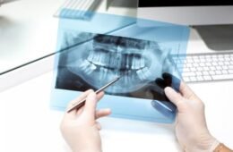
A panoramic X-ray creates a broad image of the upper and lower jaws and many of the teeth in one view. It may be useful for assessing wisdom teeth, developing teeth, jaw structures and treatment planning.
Other Dental Imaging
In some situations, dentists may recommend more advanced three-dimensional imaging, such as cone beam computed tomography (CBCT). This is not required for routine dental checkups and is generally used when more detailed information is needed for a specific diagnostic or treatment-planning purpose.
Are Dental X-Rays Safe?
Modern dental X-ray equipment uses relatively low radiation doses. Dental X-rays are generally considered safe when they are clinically justified and performed using appropriate equipment and radiation-safety procedures.
However, X-rays involve radiation, so they should not be taken simply because they are available. Dentists consider your individual circumstances and recommend imaging when the expected diagnostic benefit justifies the exposure.
How Often Do You Need Dental X-Rays?
There is no single schedule that applies to everyone. The frequency depends on factors such as your age, dental history, current oral health, symptoms, risk of tooth decay and the type of treatment being considered.
Someone with a higher risk of dental problems may need imaging at different intervals from someone with a low risk. Your dentist can recommend an appropriate schedule based on your individual needs.
Do You Need an X-Ray at Every Dental Checkup?
Not necessarily. A routine dental examination does not automatically mean that you need an X-ray every time you visit the dentist.
Your dentist decides whether imaging is useful based on your symptoms, dental history, previous X-rays and current examination findings. If there is no clinical reason for another image, an additional X-ray may not be necessary.
What Happens During a Dental X-Ray?
The process is usually quick and straightforward. The exact procedure depends on the type of X-ray being taken.
- Your dentist or dental team will explain the type of X-ray required.
- You may be asked to remove items such as jewellery or other objects that could interfere with the image.
- For an intraoral X-ray, a small image receptor may be positioned inside your mouth.
- You will be asked to remain still while the image is taken.
- The dental team will check the image to make sure it provides the information needed.
The X-ray itself generally takes only a short time. You may feel some pressure from the image receptor, but the procedure should not normally be painful.
Can Dental X-Rays Find Problems Before They Cause Pain?
Sometimes. Certain dental problems can develop without noticeable symptoms, and an X-ray may reveal changes that are not visible during a basic visual examination.
For example, decay between teeth or changes around the root of a tooth may not cause obvious symptoms in the early stages. This is one reason your dentist may recommend an X-ray even when you do not have tooth pain.
Why Are Dental X-Rays Important Before a Root Canal?
When a root canal is being considered, the dentist needs information about the affected tooth and its root structure. A suitable dental X-ray can help assess the tooth and surrounding area and support treatment planning.
The dentist will also consider your symptoms and clinical examination. The X-ray is an important diagnostic aid, but it is not used in isolation.
Why Are X-Rays Useful Before Dental Implants?
Dental implant treatment requires careful planning. Imaging may be used to assess the teeth, jawbone and nearby anatomical structures and to help determine whether additional information is required before treatment.
For some implant cases, a three-dimensional scan such as CBCT may be recommended when detailed information about bone and surrounding structures is necessary.
Are Dental X-Rays Different for Children?
Children may need dental X-rays in certain situations, but the decision is based on their individual dental development, history and risk factors. X-rays can sometimes help monitor developing teeth, detect decay or assess problems that cannot be seen clearly during a visual examination.
Because children are more sensitive to radiation than adults, dental imaging should be used only when clinically appropriate, with the lowest reasonable exposure for the required diagnostic information.
What Should You Tell Your Dentist Before an X-Ray?
Before having a dental X-ray, let your dentist know about relevant health information, especially if you are pregnant or may be pregnant. It is also useful to tell your dental team about recent dental X-rays taken elsewhere, when possible.
Sharing previous imaging may help your dentist decide whether a new X-ray is necessary.
Dental X-Rays: Key Takeaways
- Dental X-rays can show areas that cannot be fully assessed during a visual examination.
- They may help detect cavities, changes around tooth roots, bone problems and impacted teeth.
- Different types of X-rays provide different views of the teeth and jaws.
- You do not necessarily need an X-ray at every dental checkup.
- The recommended frequency depends on your individual dental needs and risk factors.
- Modern dental X-rays use relatively low radiation doses when performed appropriately.
- Tell your dentist if you are pregnant or may be pregnant.
- X-rays are used together with your symptoms and clinical examination to make a diagnosis.
Frequently Asked Questions
Your dentist may recommend an X-ray to examine areas that cannot be seen clearly during a routine visual examination, such as between teeth, around tooth roots or within the jawbone.
Dental X-rays are generally not painful. Some intraoral X-rays may cause temporary pressure or mild discomfort when the image receptor is positioned inside the mouth.
Dental X-rays use relatively low amounts of radiation and are generally considered safe when clinically justified and performed using appropriate radiation-safety procedures.
No. Your dentist decides whether an X-ray is needed based on your symptoms, dental history, oral health, previous imaging and individual risk factors.
Yes, certain dental X-rays can help identify decay in areas that are difficult to see during a visual examination, including some cavities between teeth.
An X-ray can sometimes show changes around the root of a tooth that may be associated with infection. Your dentist will consider the X-ray together with your symptoms and clinical examination.
If you are pregnant or think you may be pregnant, tell your dentist before an X-ray. Your dental team can determine whether imaging is necessary and take appropriate precautions.
A panoramic X-ray provides a broad view of the upper and lower jaws and many of the teeth in one image. It can be useful for assessing wisdom teeth, developing teeth and certain jaw structures.
Need a Dental Checkup?
A dental examination can help identify problems early and determine whether an X-ray or other diagnostic test is appropriate for your situation.
Book a Dental Appointment